Checklist dataset Registered 5 October 2023
Revision of the huntsman spider genus Micrommata Latreille, 1804 (Sparassidae Sparassinae)
Published by Plazi.org taxonomic treatments database
Genus
Micrommata Latreille, 1804
Classification and descendants
Loading
Loading
Loading
Loading
Loading
Loading
Media
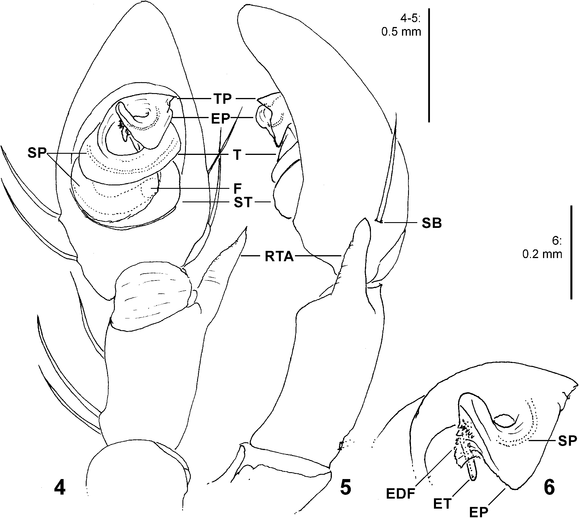
FIGURES 4–6. Micrommata aljibica Urones, 2004, holotype male, left palp (4 ventral, 5 retrolateral, 6 tegular tip ventral). EDF—embolic denticle field, EP—embolic plate, ET—embolus tip, F—fundus of spermophore, RTA—retrolateral tibial apophysis, SB—cymbial spine base, SP—spermophor, ST—subtegulum, T—tegulum, TP—tegular prong.
Jäger, Peter
CC_BY
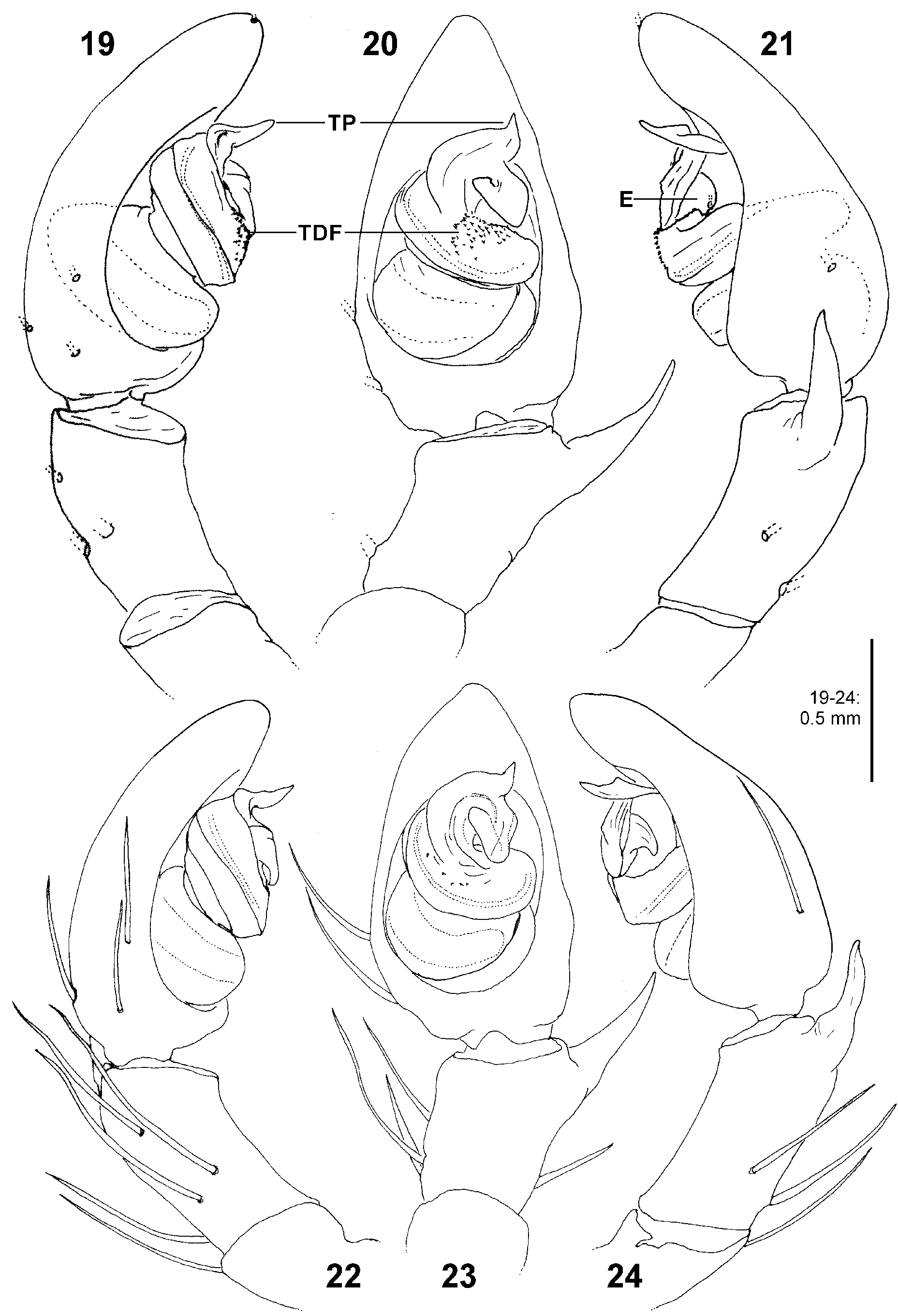
FIGURES 19–24. Micrommata biggi spec. nov., left palp (19, 22 prolateral; 20, 23 ventral; 21, 24 retrolateral) (19–21 holotype male from Ţrkiye; 22–24 male from Turkmenistan). E—embolus, TDF—tegular denticle field, TP—tegular prong.
Jäger, Peter
CC_BY
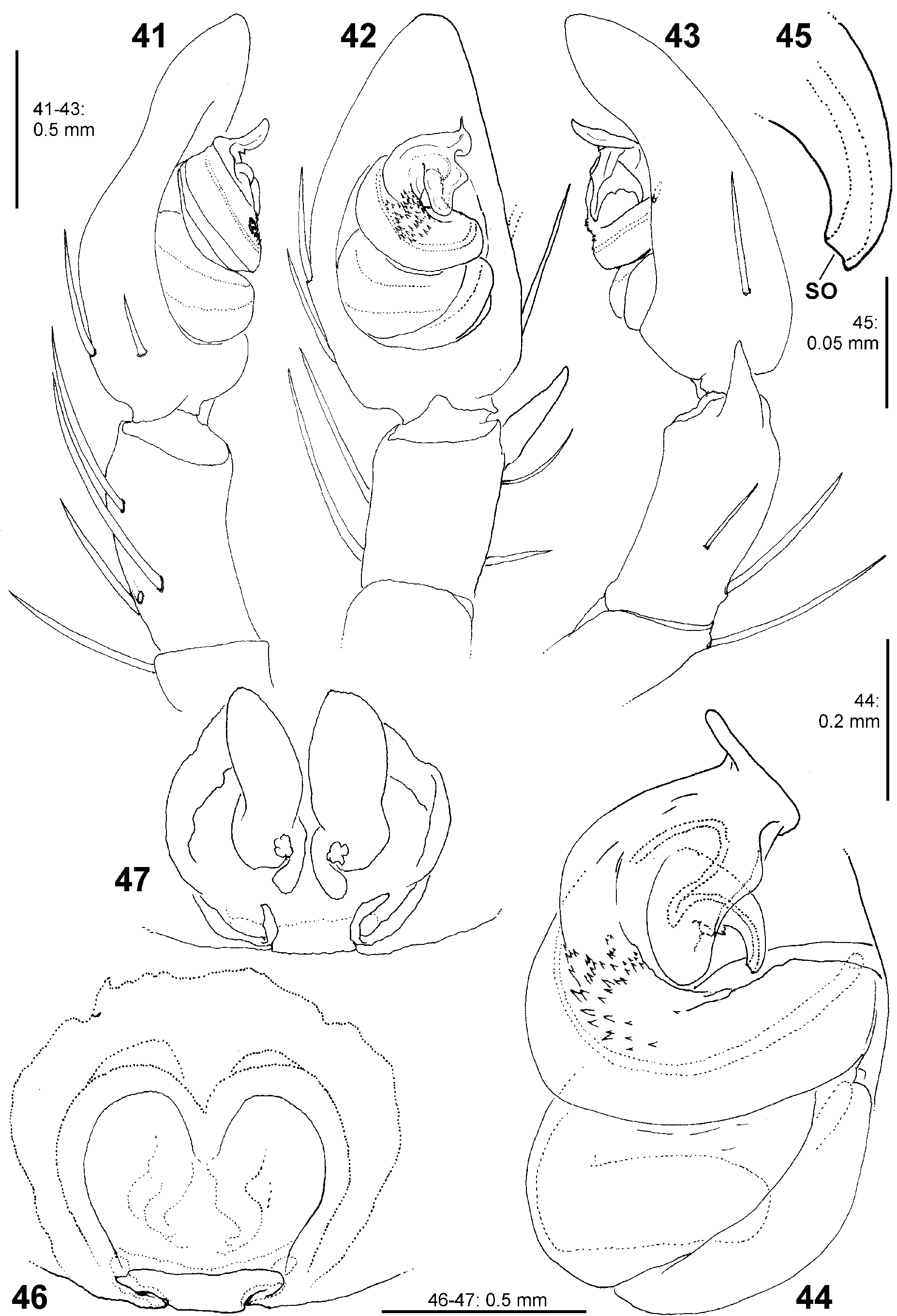
FIGURES 41–47. Micrommata diesenhoff spec. nov. from Sierra Leone, copulatory organs. 41–45 Left palp (41 prolateral; 42 ventral; 43 retrolateral; 44 tegulum, ventral; 45 embolus tip ventral). 46–47 Female copulatory organ (46 epigyne, ventral; 47 vulva dorsal) (41–45 holotype male; 46–47 female paratype). SO—spermophor opening.
Jäger, Peter
CC_BY
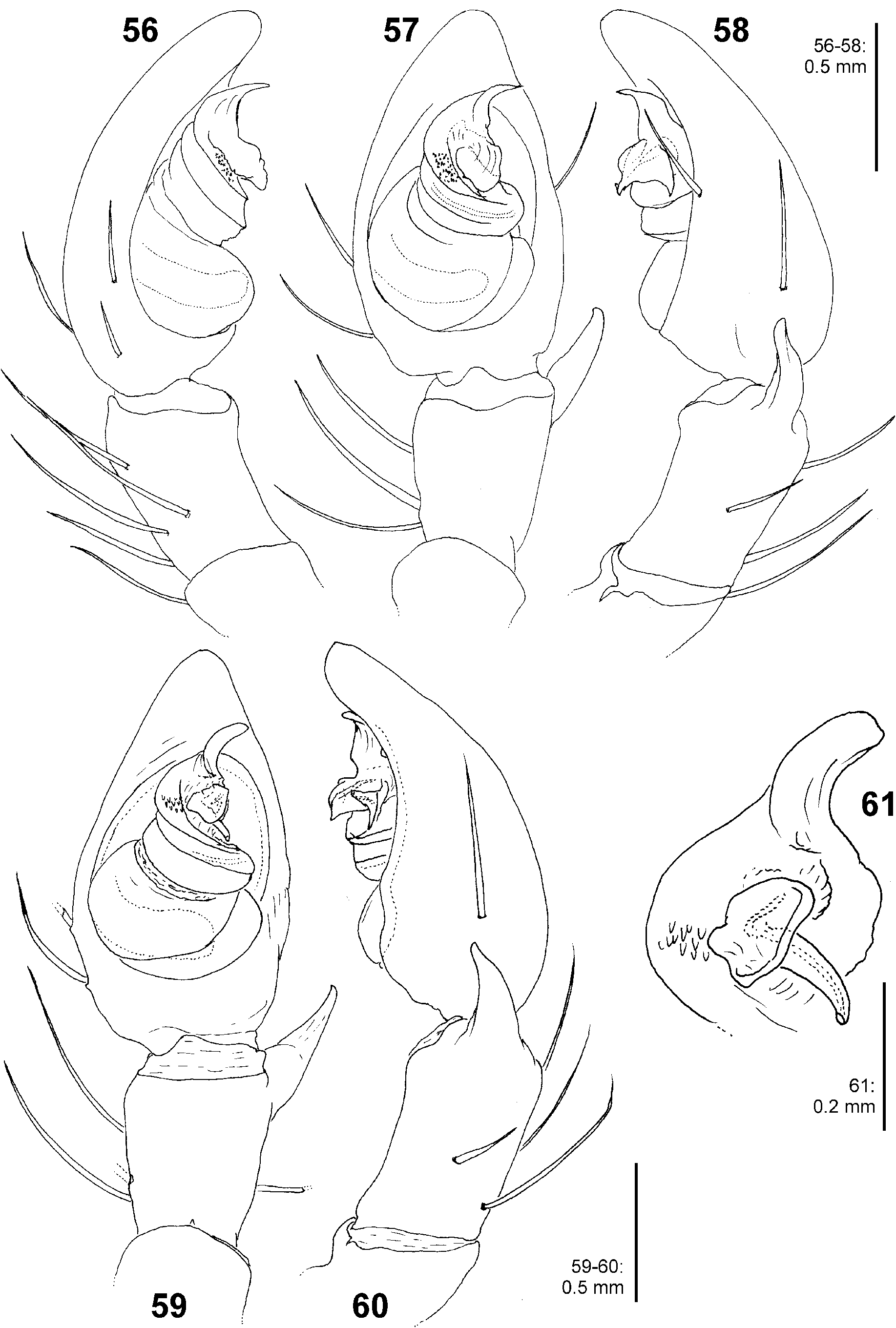
FIGURES 56–61. Micrommata formosa, left palp (56 prolateral; 57, 59 ventral; 58, 60 retrolateral; 61 tegulum tip and embolic division, ventral) (56–58 from Israel; 59–61 male from Spain).
Jäger, Peter
CC_BY

FIGURES 84–89. Micrommata ligurina, male. 84 Left palp, ventral. 85 Ditto, retrolateral. 86–87 leg claws, lateral.88 Trilobate membrane, dorsal. 89 Prosoma with eye arrangement, frontal (84–85 from Spain; 86–89 from Italy: Sardinia). BO—basal outgrowth of embolus, DI—incision of distal tegular margin.
Jäger, Peter
CC_BY
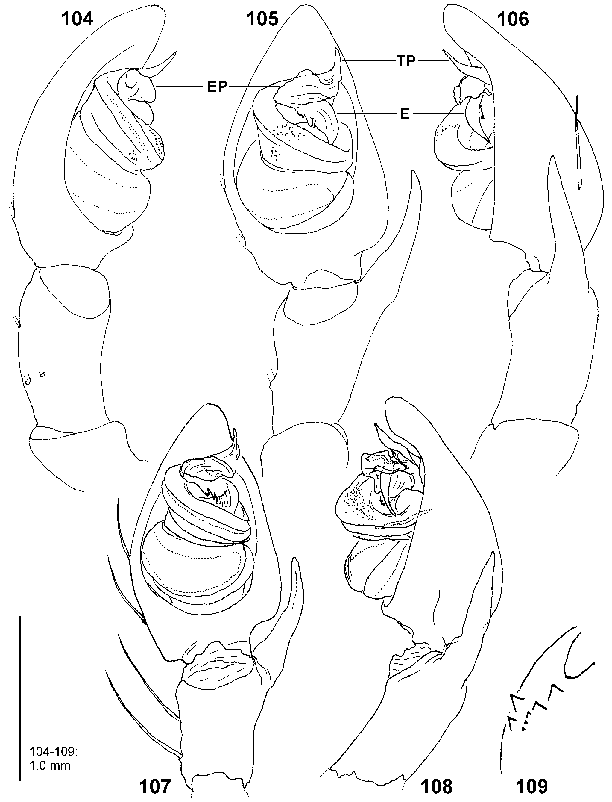
FIGURES 104–109. Micrommata virescens, male. 104–108 Left palp (104 prolateral; 105, 107 ventral; 106, 108 retrolateral). 109 Left chelicera, ventral (104–106 from Russia; 107–108 from Japan; 109 from Germany). E—embolus, EP—embolic plate, TP—tegular prong.
Jäger, Peter
CC_BY
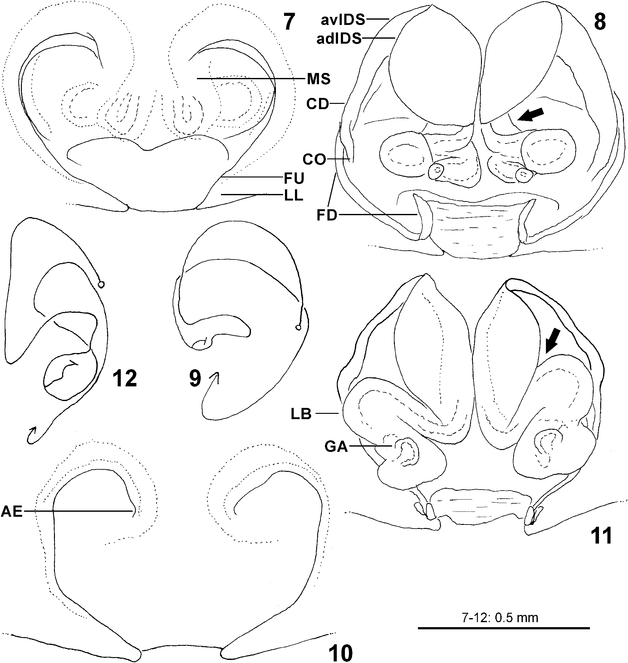
FIGURES 7–12. Micrommata aljibica Urones, 2004, females, copulatory organs (7, 10 epigyne, ventral; 8, 11 vulva dorsal; 9, 12 schematic course of IDS, dorsal) (7–9 paratype, 10–12 MNCN 20.02/19884). adIDS—antero-dorsal part of internal duct system, AE—antero-median ends of epigynal furrows, avIDS—antero-ventral part of internal duct system, CD—copulatory duct, CO—copulatory opening, FD—fertilisation duct, FU—epigynal furrow, GA—glandular appendages, LB—lateral bulge of internal duct system, LL—lateral lobes, MS—median septum; arrows indicating the incision between anterior and posterior part of IDS.
Jäger, Peter
CC_BY
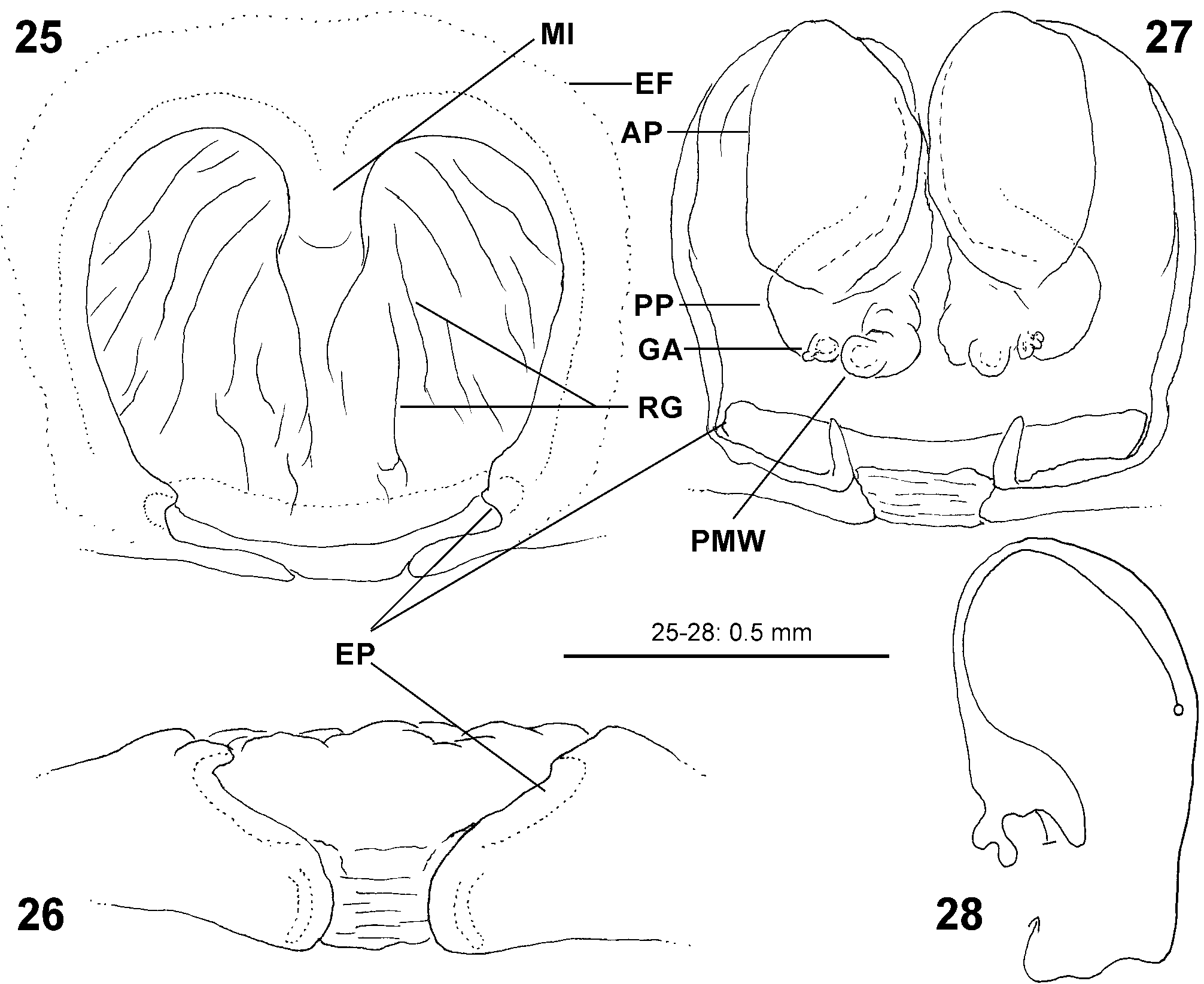
FIGURES 25–28. Micrommata biggi spec. nov., female from Iran, copulatory organ (25 epigyne, ventral; 26 ditto, posterior; 27 vulva dorsal; 28 schematic course of IDS, dorsal). AP—anterior part of internal duct system, EF—epigynal field, EP—epigynal pocket, GA—glandular appendages, MI—antero-median incision of median septum, PMW—postero-median windings, PP— posterior part of internal duct system, RG—radial grooves of median septum.
Jäger, Peter
CC_BY
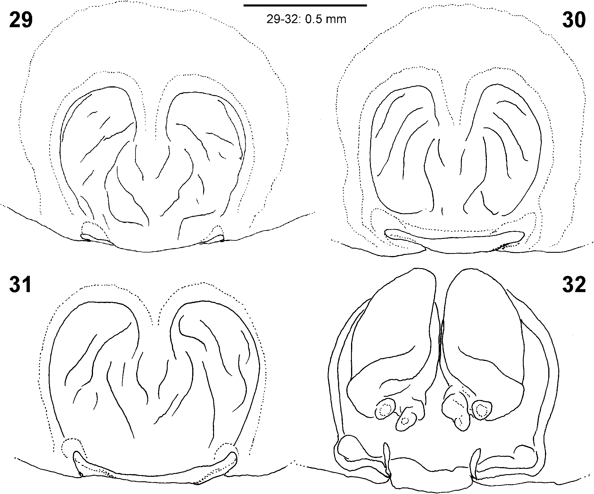
FIGURES 29–32. Micrommata biggi spec. nov., females from Turkmenistan, copulatory organ (29–31 epigyne, ventral; 32 vulva dorsal).
Jäger, Peter
CC_BY
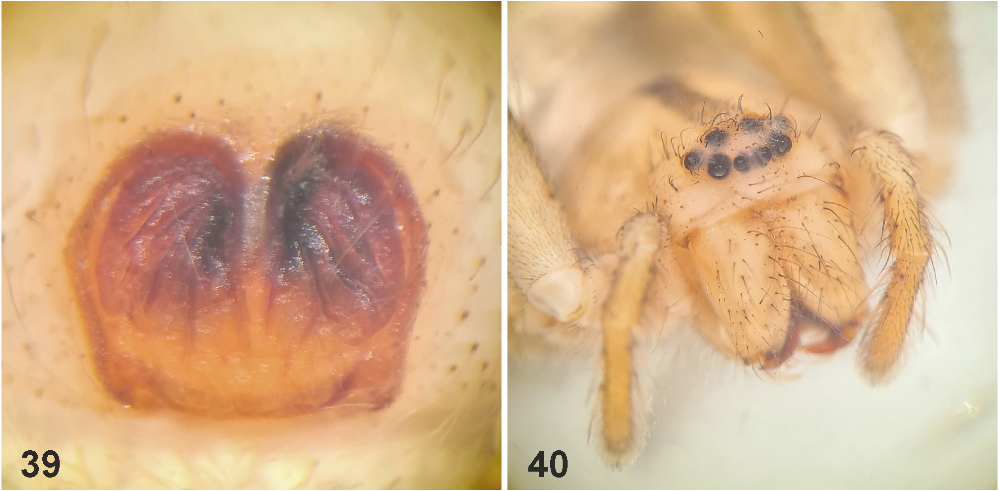
FIGURES 39–40. Micrommata biggi spec. nov., female paratype from Ţrkiye. 39 Epigyne, ventral. Habitus, frontal.
Jäger, Peter
CC_BY

FIGURES 48–53. Micrommata diesenhoff spec. nov., female paratypes from Sierra Leone, copulatory organs (48, 51 epigyne, ventral; 49, 53 vulva dorsal; 50 schematic course of IDS, dorsal; 52 epigyne, posterior). SS—slit sensillum.
Jäger, Peter
CC_BY
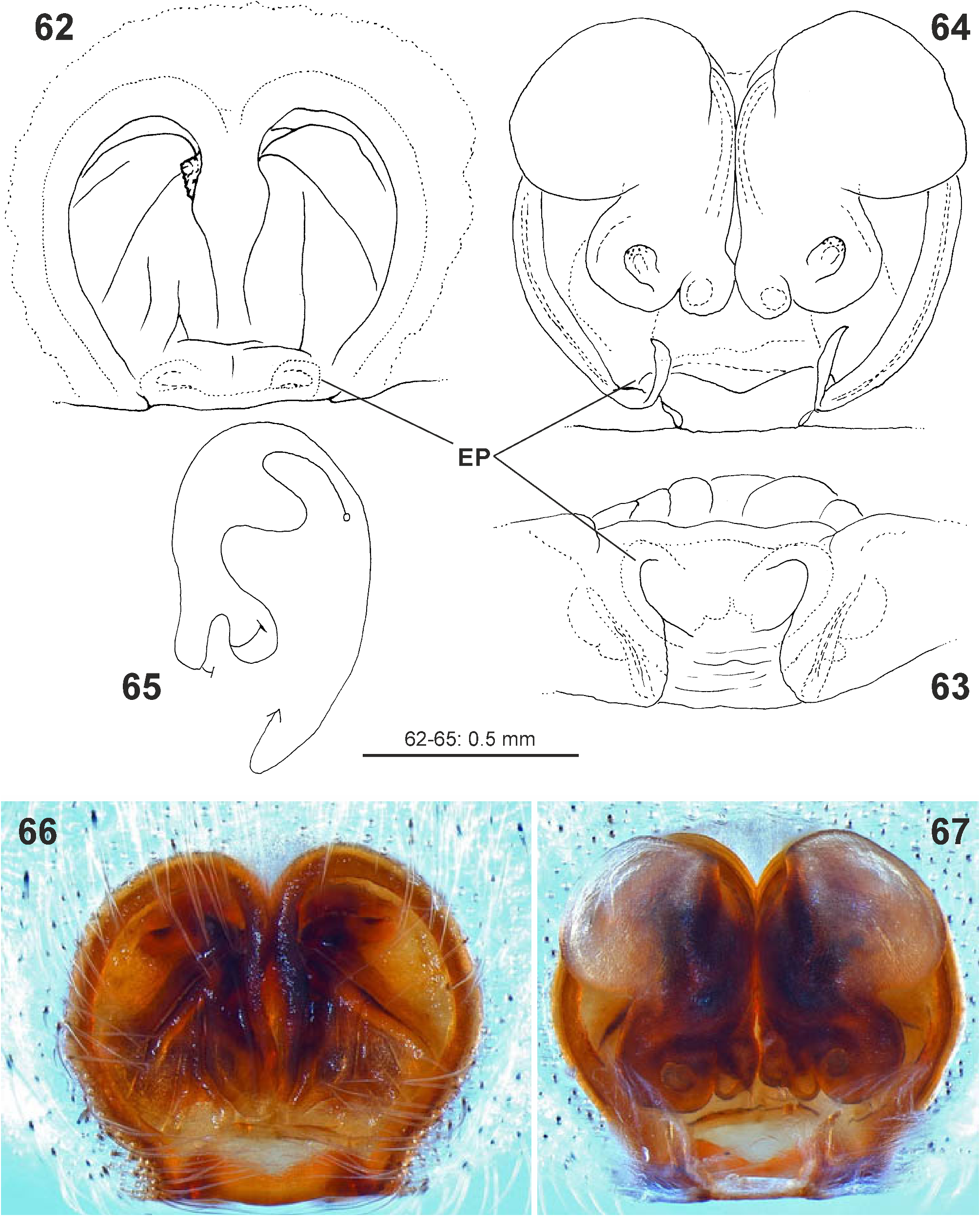
FIGURES 62–67. Micrommata formosa, female copulatory organ (62, 66 epigyne, ventral; 63 ditto, posterior; 64, 67 vulva, dorsal; 65 schematic course of IDS, dorsal) (62–65 holotype female of M. aragonensis from Spain; 66–67 female from Spain); photo credits: 66–67 Arnaud Henrard. EP—epigynal pocket.
Jäger, Peter
CC_BY
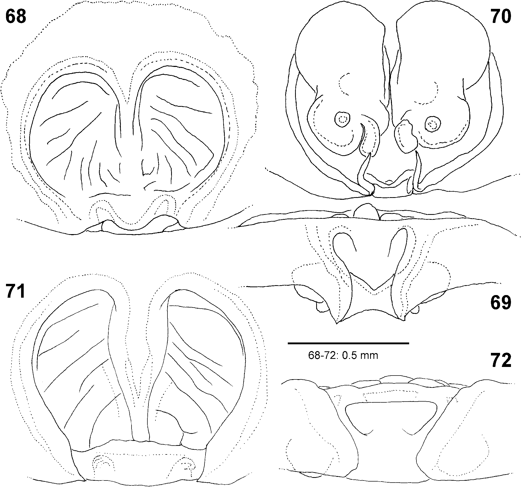
FIGURES 68–72. Micrommata formosa, female copulatory organ (68, 71 epigyne, ventral; 69, 72 ditto, posterior; 70 vulva, dorsal (68–70 from Israel; 71–72 from Tunisia).
Jäger, Peter
CC_BY
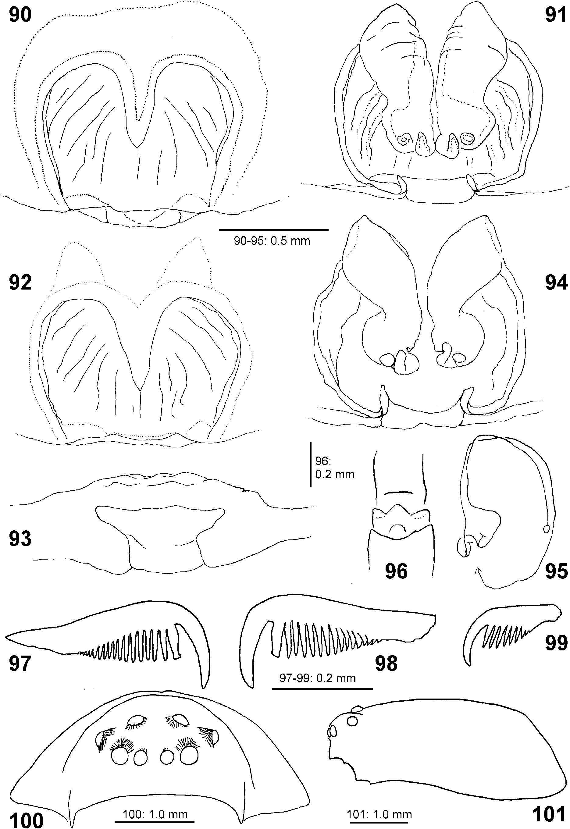
FIGURES 90–101. Micrommata ligurina, female. 90–95 Copulatory organ (90, 92 epigyne, ventral; 91, 94 vulva, dorsal; 93 epigyne, posterior; 95 schematic course of IDS, dorsal). 96 Trilobate membrane, dorsal. 97–98 Leg claws, lateral. 99 Palpal claw, lateral. 100–101 Prosoma with eye arrangement (100 frontal; 101 lateral) (90–95 from Spain; 96–101 from Italy: Sardinia).
Jäger, Peter
CC_BY
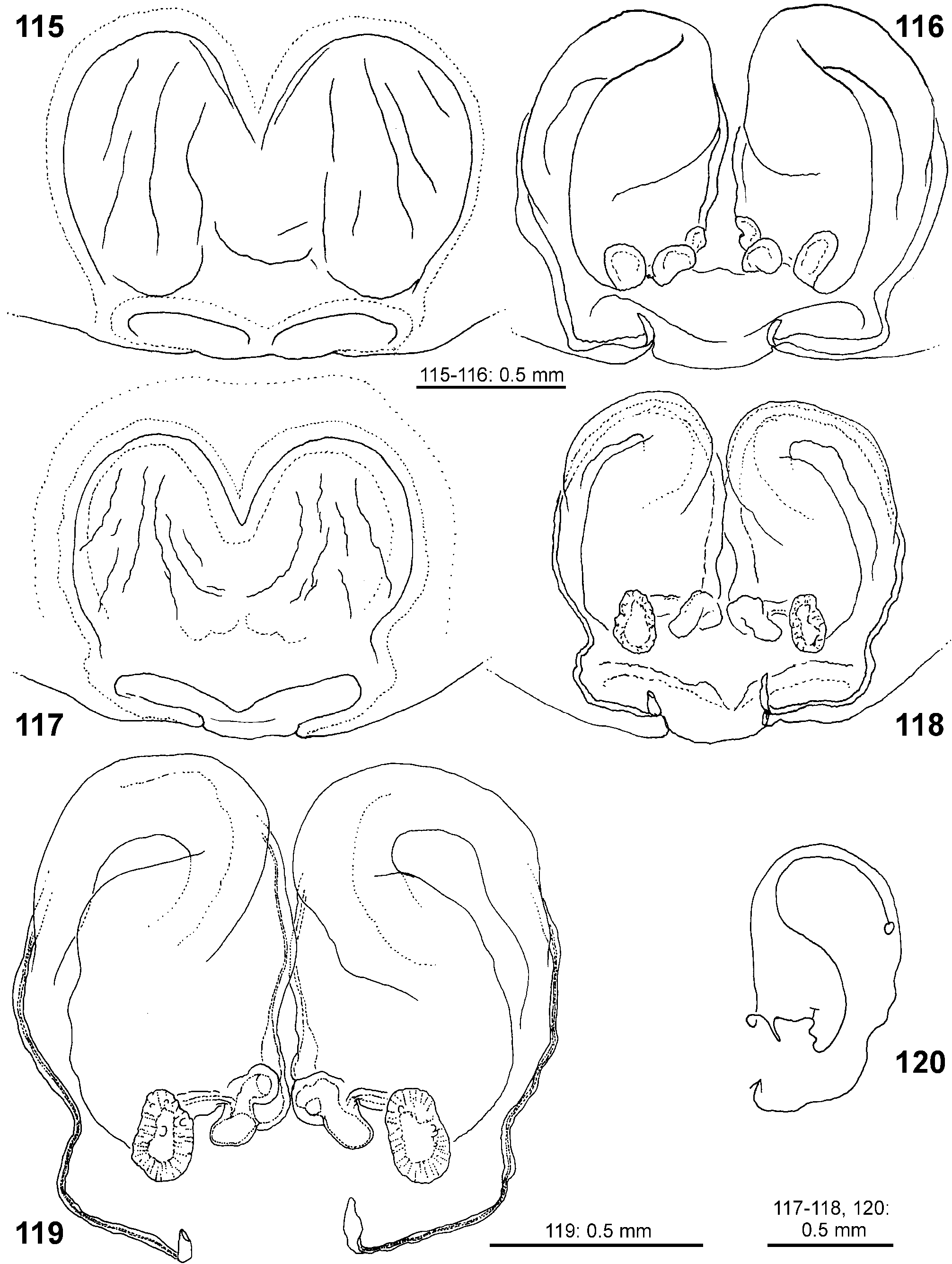
FIGURES 115–120. Micrommata virescens, female copulatory organ (115, 117 epigyne, ventral; 116, 118–119 vulva, dorsal; 120 schematic course of IDS, dorsal) (115–116 from Russia; 117–120 from Japan).
Jäger, Peter
CC_BY
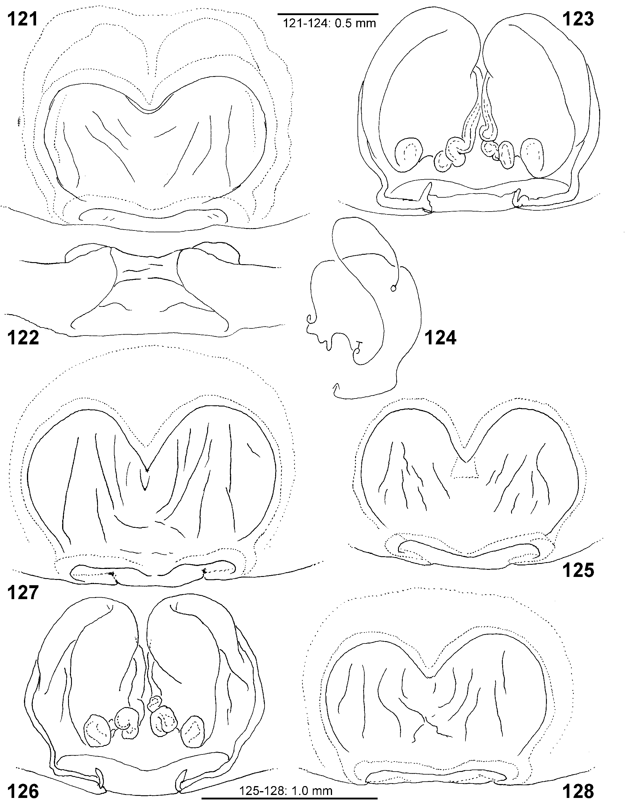
FIGURES 121–128. Micrommata virescens, female copulatory organ (121, 125 127–128 epigyne, ventral; 122 ditto, posterior; 123, 126 vulva, dorsal; 124 schematic course of IDS, dorsal) (121–124 from Germany; 125–127 from Kazakhstan; 128 from Russia).
Jäger, Peter
CC_BY
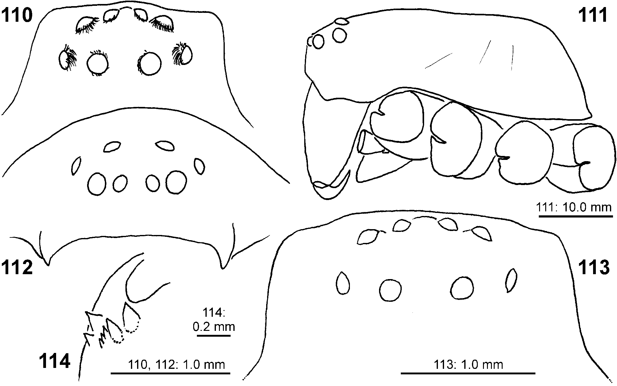
FIGURES 110–114. Micrommata virescens from Germany. 110–113 Prosoma with eye arrangement (110, 113 dorsal; 111 lateral; 112 frontal). 114 Left chelicera, ventral (110–112 male; 113–114 female).
Jäger, Peter
CC_BY
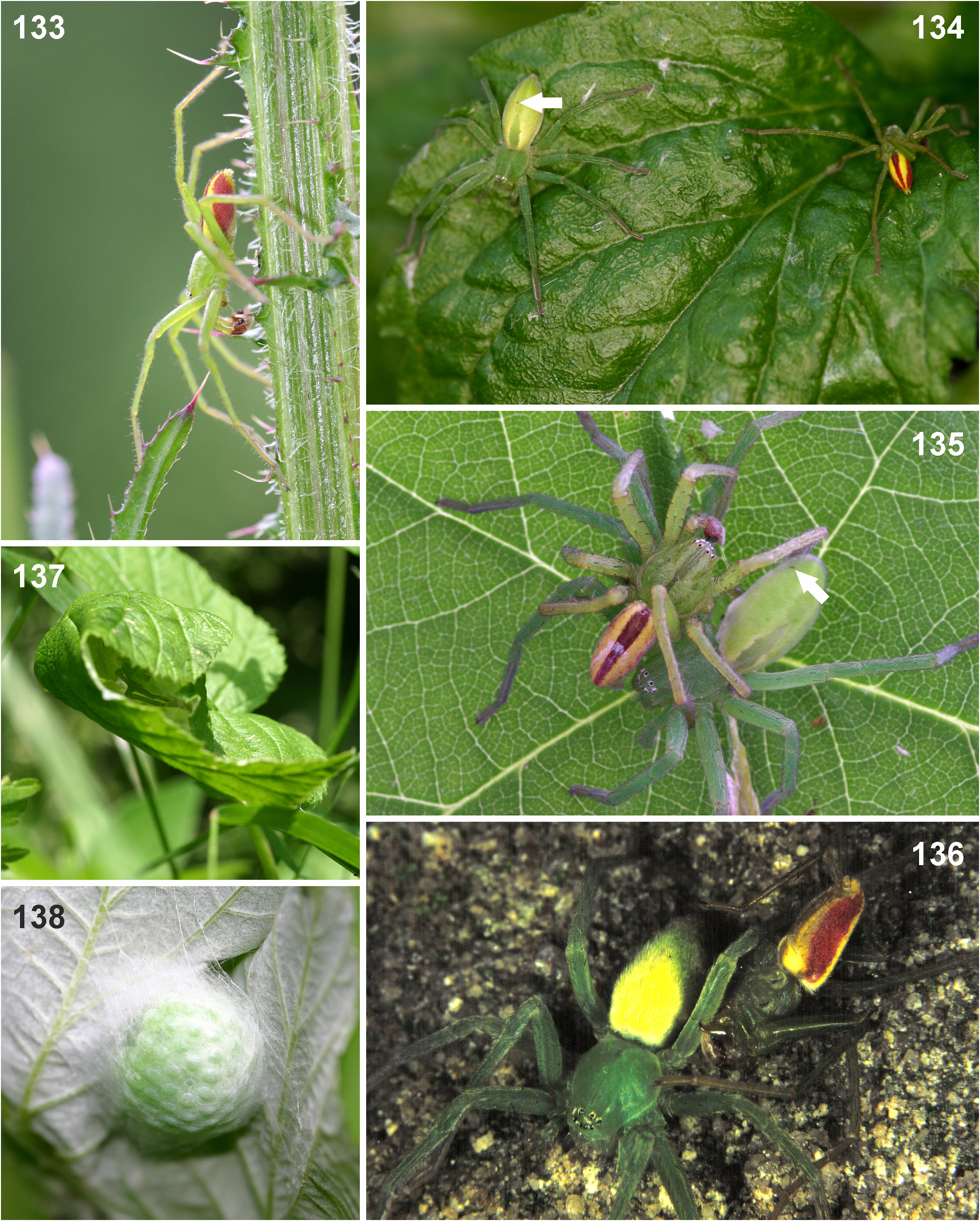
FIGURES 133–138. Micrommata virescens, adults from Germany, habitus and behaviour. 133 Male, looking for a female. 134 Female and male in the natural habitat. 135 Male and female in mating position. 136 Ditto, male bites leg of female. 137 Female with egg sac in rolled leaf of Rubus sp. 138 Egg sac with green eggs. Arrows indicating scars on the opisthosoma of female spiders. Photo credit: 136 Herbert Schirmer.
Jäger, Peter
CC_BY
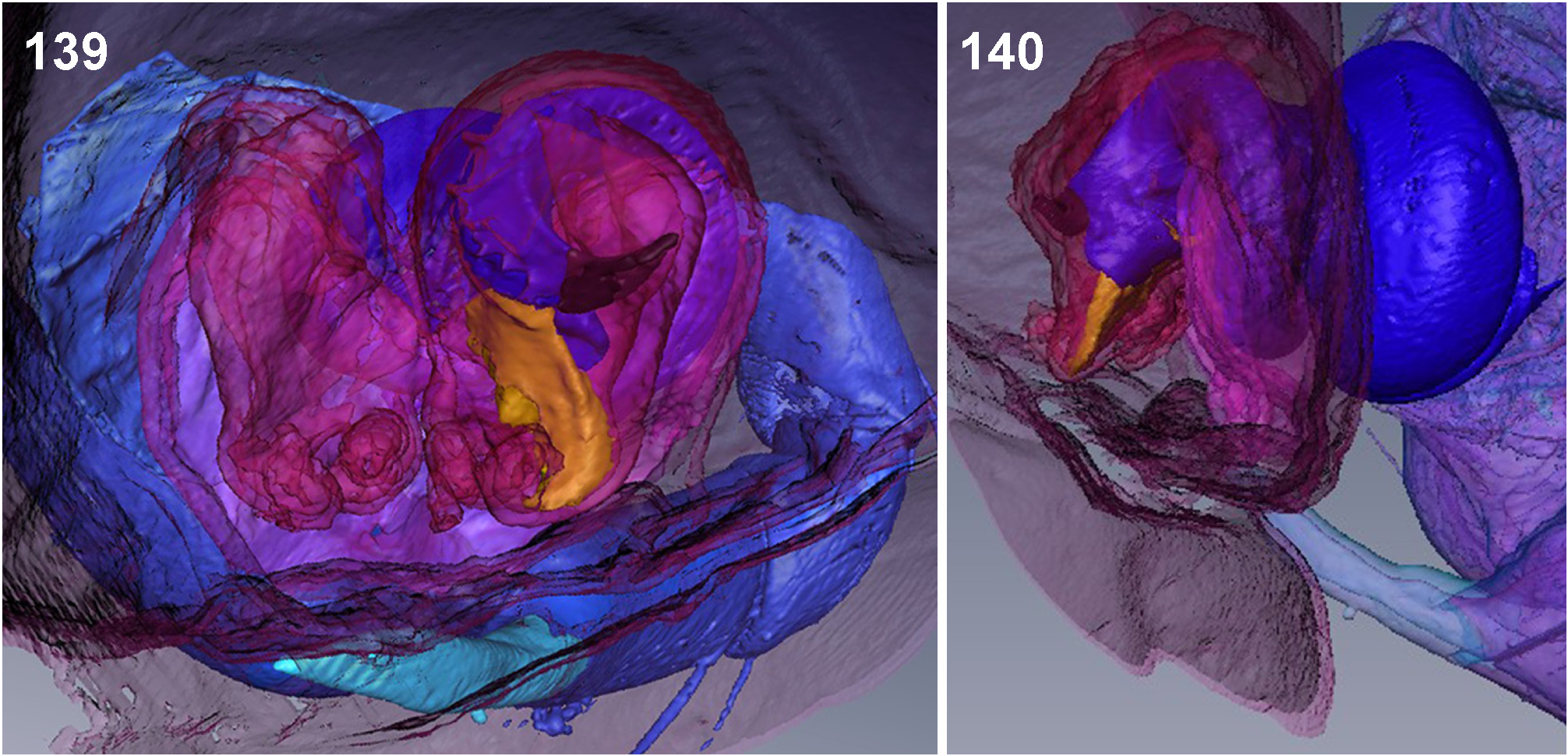
FIGURES 139–140. Micrommata virescens, µ-CT pictures of male and female genitalia during copulation (139 dorsal; 140 lateral). Pink: female internal duct system; light blue: RTA; light violet: cymbium plus basal haematodocha; dark blue: subtegulum and tegulum; black: tegular prong; golden: embolic division.
Jäger, Peter
CC_BY
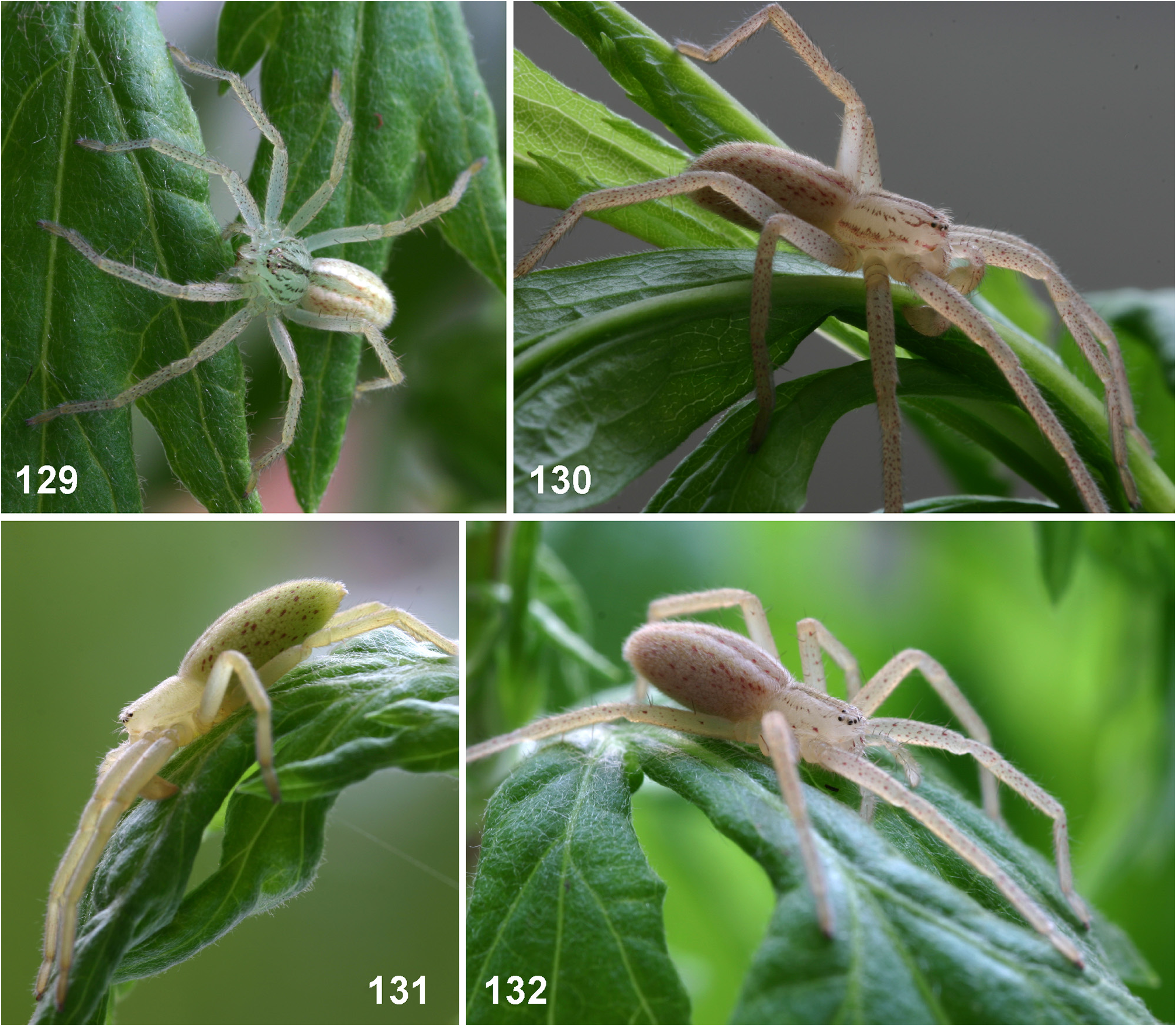
FIGURES 129–132. Micrommata virescens, immatures from Germany, habitus (129, 132 juvenile; 130–131 subadult male).
Jäger, Peter
CC_BY
Bibliography
1 result
Latreille, P. A. (1804) Tableau methodique des Insectes. Nouveau Dictionnaire d'Histoire Naturelle, Paris, 24, 129 - 295. |
Citation
Micrommata Latreille, 1804 in undefined